Institute of Physics
Maria Curie-Sklodowska University
Lublin, Poland
Institute of Physics
Maria Curie-Sklodowska University
Lublin, Poland
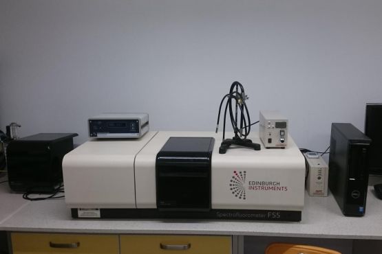
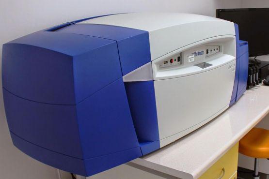
Edinburgh Instruments FS5
Our Edinburgh Instruments FS5 spectrofluorometer is allows various types of fluorescence measurements with simultaneous monitoring of absorption and dynamic correction. It allows us to perform low temperature analysis (down to 77K) in the spectral range of 250-1650 nm. Recently we were able to make an upgrade the system to allow excitation and emission time resolved measurements in the range of 10 µs - 10 s. We have a full range of sample compartments for various samples.
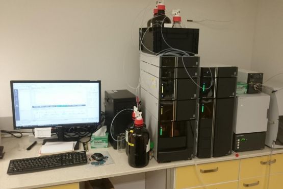
Nexera
Our lab is equiped with two Shimadzu Nexera HPLC systems.
- A binary pump system with a PDA detector and a manual injection port.
- A dual LC-40D XR pump system (binary and quaternary LPG), SIL-40C XR autosampler, flow-line selection valve, PDA detector (SPD-M40) followed by an ELSD. We can also automate fraction collection with an FRC-10A with input from the detector of choice.
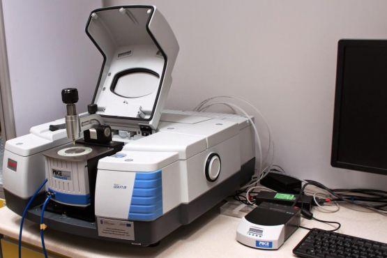
FTIR Nicolet iS50
Our Fourier Transform Infrared Spectroscope is equipped in a DLaTGS and MCT detector, ZnSe and Ge crystals and a diamond ATR. We have the possibility to carry out ramped temperature measurements both in the multiple and single reflectance modes.
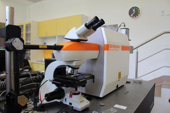
Renishaw
Raman spectroscopy is a based on inelastic scattering of light. Combining this spectroscopic technique with a confocal microscope allows us to gather data in any point of the microscopic sample creating a Raman map depicting a variation in spectral information in each pixel. Our system is combined with an Atomic Force Microscope (AFM) that allows us to additionally image the topography of soft biological materials in their native environments as well models of lipid membranes.
Note: Our JPK AFM is interchangeable between the Raman imaging and FLIM systems, giving us great flexibility in analyzing microscopic structures in the nanometer scale.
The Renishaw system is equipped in four laser lines (457,9 nm, 488 nm, 514 nm, 633 nm); 200-1000 nm CCD camera of a 0,5 cm-1 and a high-sensitivity EMCCD camera (dark current 0,00007 e-/pixel/s).
Chirascan Plus
A Circular Dichroism (CD) spectrometer is used to measure circularly polarized light. CD spectroscopy is very valuable method in the analysis of structural properties of molecular systems and changes in their organization.
Our Chirascan Plus spectrometer from Applied Photophysics is equipped in a avalanche photodiode detector, xenon lamp (165-1150 nm) and allows us to measure sample from -20 °C to 80 °C (even at 77K when combined with a Oxford DN2 cryostat).
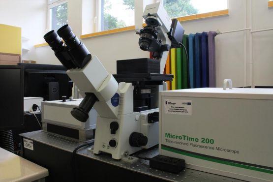
Picoquant MicroTime MT200
We are very luck to work with one of the best set ups in Poland. Fluorescence Lifetime Imaging Microscopy (FLIM) is a method that is based on the measurement on the fluorescence lifetime in conjunction with a confocal microscope. FLIM measures parameters of fluorescence such as intensity, wavelength, lifetime and polarization in any given point of a microscopic sample. It can provide information about dynamic processes occurring in the sample, molecular organization of species of interest, interaction between molecules and their orientation.
The system consists of a Picoquant MicroTime 200 main module, Olympus IX71 confocal microscope; six pulse lasers, including a tunable femtosecond 690 – 1020 nm 4W laser; six picosecond diode-pumped solid-state lasers (~1 mW): 375 nm, 405 nm, 440 nm, 470 nm, 510 nm, 640 nm; two Picoquant τ-SPAD photon counting detectors (350 ps resolution); PMA hybrid detector (120 ps resolution).